From Blindness to Brightness: Sahyadri Narayana Doctors perform complicated craniotomy, removes tumour


Shivamogga, June 07 : What started as a mild blurriness in her right eye soon turned into complete darkness for a 47-year-old woman. At first, she brushed it off as a minor issue — perhaps it was just age catching up. But within days, her thoughts turned into fear as she lost all vision in that eye, unable to even perceive light. Little did she know, it wasn’t an eye problem at all, but a large brain tumour growing silently behind her eye, threatening not just her sight but her very life.
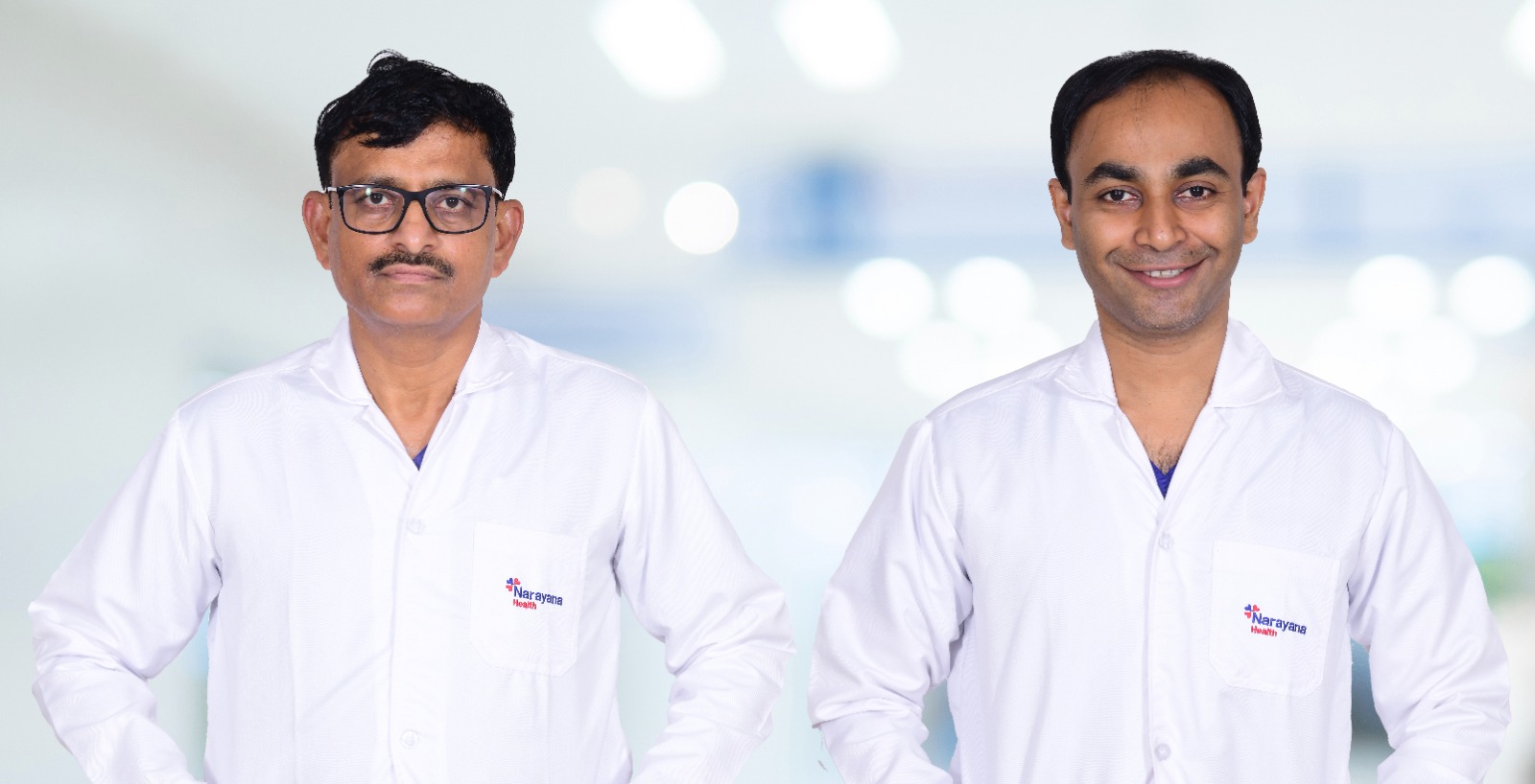
She journeyed to Sahyadri Narayana Multispeciality Hospital in Shivamogga, where a team led by neurosurgeon Dr. Anilkumar M.S discovered a massive brain tumour dangerously pressing on her optic nerve and major blood vessels in the brain. Thanks to a marathon 12-hour surgery led by the neurosurgeon, the woman has now regained vision — and her life.
The woman, whose identity is being withheld on request for privacy, arrived at the hospital after experiencing complete blindness in her right eye for two weeks. “She couldn’t even see light with that eye. The vision loss was gradual at first but progressed rapidly,” Dr Anilkumar said.
Giving details of the case, Dr Anilkumar explained that an MRI scan revealed the startling cause – a right medial sphenoid wing meningioma, which is a large, slow-growing brain tumour settled deep inside the skull. Measuring approximately 5 x 4 x 3.5 cm, the tumour had silently grown to encase the right optic nerve and was dangerously close to the middle cerebral artery (MCA), a crucial blood vessel in the brain.

Doctors feared that the tumour would extend further, potentially compressing the hypothalamus which is a critical area that regulates consciousness and body functions. Left untreated, the patient would be risking coma or even death.
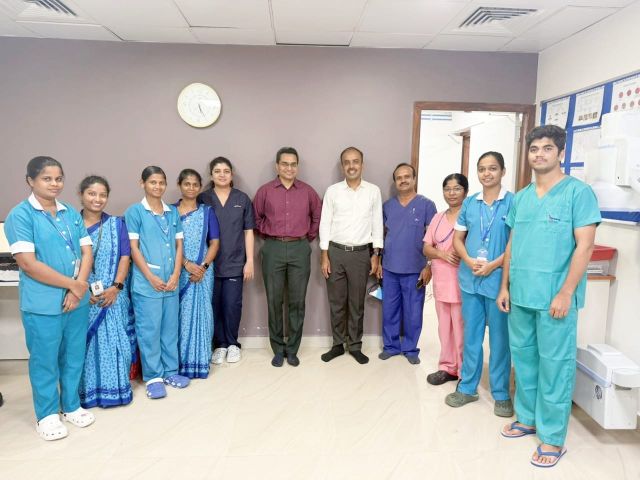
Despite the grim diagnosis, the neurosurgical team, led by Dr. Anilkumar M.S, took up the challenge. The procedure — a delicate craniotomy aided by advanced microscopic and neuronavigation technology — took nearly 12 hours. Using high-precision tools, the team carefully peeled the tumour off the optic nerve and surrounding structures, millimetre by millimetre.
“The location was extremely sensitive. We were navigating near vital centres for vision, memory, and consciousness. One wrong move could have caused permanent damage,” explained Dr. Anilkumar.
What makes this story remarkable is the outcome. Just two weeks after surgery, the patient shocked even her doctors — she could count fingers from six feet away using the same eye that had gone completely blind. “In most cases where the optic nerve is already encased, chances of recovery are minimal. But this patient beat the odds,” said Dr Anilkumar.
Visual recovery in such cases is considered rare. According to global studies, only five to eight per cent of patients with medial sphenoid wing meningiomas involving the optic nerve regain any vision after surgery. In cases like this, deterioration is more common, occurring in 22–35 per cent of patients.
Beyond vision, doctors were also concerned about the tumour’s proximity to areas responsible for memory and hormonal control. Had it progressed further, the tumour might have compressed the hypothalamus, leading to memory loss, hormonal imbalance, or even coma.
Thanks to timely intervention, not only was the woman’s vision partially restored, but her brain functions were also preserved. She was discharged in a stable condition with follow-up care underway.
This case is also a wake-up call for many who ignore gradual vision changes, and headaches mistaking them for age-related decline. As Dr Anilkumar warned, “Sudden or gradual loss of vision and headache should never be ignored. Sometimes, the eye is not the problem. It’s the brain.”